

When I opened my current practice in 2015, I knew I would be diagnosing and treating dry eye disease (DED), but I did not intend it to be my main focus. Within a year, however, 29 doctors were referring their patients to me for DED treatment, and today, about 90% of my practice is devoted to DED.
Looking back after that first year, I wondered how I had become the go-to doctor for DED. Certainly it was related to outcomes, but I was not doing anything magical. Patients were achieving excellent outcomes because they were adhering to the treatments I prescribed, and this compliance tracked back to engaging and educating these patients. Once they could see and understand what was happening in their eyes and believed that I could help them, they became invested in their treatment.
Streamlined Dry Eye Evaluation
There’s no question that DED is complicated. We have abundant data on the mechanisms at play, and we have numerous therapies to treat the various manifestations of this disease. We also have sophisticated diagnostic and imaging equipment to help us identify the causes and effects of DED. What has been lacking is an efficient method for performing evaluations and educating patients in a way that will capture their attention and encourage compliance without disrupting the workflow of the office.
The cornerstone of my practice, the Keratograph 5M (Oculus) provides data from a battery of tests to aid in my diagnoses and treatment decisions, and it helps me explain my findings and recommendations to patients.
The newest feature of the Keratograph 5M, the Crystal TEAR Report, is the result of my 18-month collaboration with the engineers of the Oculus R&D team in Germany. Our goal was to streamline the entire DED evaluation process from image capture to data assessment to patient education.
The Keratograph 5M performs all the relevant tests for a full DED evaluation, and the new software ensures that those tests are performed quickly and in logical order to produce good quality scans (measuring water before performing tear-inducing tests, for example). The program is fully customizable, so that practitioners can choose which tests will be performed, depending on the type of visit (new patient evaluation, follow-up, screening), and they can preset their preferred therapies for each type and stage of DED. A one-time commitment to inputting these preferences produces a robust, time-saving program.
Of particular importance to me is the ability to create a collage of nine images that represent the key findings I want to discuss with a patient. This is how I tell each patient’s story.
Compelling Patient Education
At my initial appointment with a new patient referred to me for a DED evaluation, I say, “We know you have dry eye. Today, we will do something different. We will find out why.”
After acquiring all the relevant data provided by the Keratograph 5M, a process that takes my technician about 10 minutes, I perform my assessment. Then, I display the collage of images and explain my findings to the patient, pointing out the abnormalities and deficiencies that are causing his or her symptoms.
The Crystal TEAR Report includes a color-coded pie chart that shows patients (and me) at a glance their status in the categories of water, oil, allergy, inflammation, bacteria, lid function, and systemic and environmental issues (Figure). This shows patients the gravity of specific issues and helps me prioritize treatments.
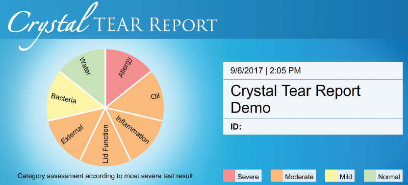
Figure. The Crystal TEAR Report includes a color-coded pie chart.
In about 3 minutes I can tell patients their story, and when I am done I can see on their faces a palpable hope that they have never had. They have been to doctor after doctor. They have been told to use artificial tears a hundred times, but they know that does not work. Finally, they feel like today is ground zero, that something will be different this time. All of a sudden they understand. The Crystal TEAR Report motivates patients to follow the treatment plan created just for them. When they start experiencing positive results, their perspective changes completely, and their confidence and trust in their doctor grows.
Positive Outcomes Via Education
I know what it looks like when a patient’s DED is treated at year 1 instead of year 2, or year 2 instead of year 6. I know what it means to his or her outcomes, and I know what it takes to make that happen. Successfully treating DED takes seeing it and, more importantly, being able to show patients the abnormalities and deficiencies that are causing their symptoms. My hope is that doctors who were reluctant to tackle DED will recognize that they can now implement an efficient and effective management program that will change their patients’ lives.

*The Dry Eye Institute is an all-day, hands-on workshop led by Dr. Brimer at her practice in Wilmington, North Carolina. With a class limit of five doctors, the goal is to create a customized plan for each attendee who will receive a kit that contains all of the tools necessary for successful implementation.