For patients and practitioners alike, visual field testing is not a favorite among diagnostic tests. From the patient’s perspective, it’s no wonder, as the test requires the patient’s attention and alertness for several minutes per eye while sitting in a dark room with a patch over the other eye.
From the practitioner’s perspective, the variability and frequent unreliability of this subjective test can lead to even further frustration when it has to be repeated in order to confirm a result. Also consider that most glaucoma patients will face many years of testing over the course of their disease and life, and visual field testing seems pretty daunting.
On the other side of the diagnostic spectrum we now have OCT. With the continued development of this technology and its expansion into general eye care practice, plus its fast, noninvasive assessment of the eye, it’s easy to see why visual field testing often takes a back seat in the diagnostic lineup for glaucoma suspects and even patients with glaucoma. The sensitivity, speed, reliability, and objective nature of OCT gives it several advantages over visual field testing.
And there are other new diagnostic tests for glaucoma (eg, electrodiagnostics and corneal hysteresis) that offer additional information to supplement visual field testing. That being said, the case for continuing to perform visual field testing on our glaucoma patients is strong. Let’s look at some of the top reasons to keep it in our repertoire.
REASONS FOR USING VISUAL FIELD TESTING
Standard of Care
In published clinical care guidelines for optometry and in consensus standards of practice, visual field testing is identified as a key component in the workup and evaluation of patients with glaucoma.1 Deviating from this is not consistent with community standard.
Assessment of Disease Severity
Traditional assessment of glaucoma disease severity is frequently made based solely upon visual field test results. Although a more detailed assessment could include optic nerve cupping and retinal nerve fiber layer analysis, the current ICD-10 diagnostic coding system, which includes a disease severity designation, is based solely on the patient’s visual field test results in the worse eye.
In 2011, a working group of the American Glaucoma Society developed a new staging system that was incorporated into the ICD-10 system in 2013.2 This makes it one of the most frequently used staging schemes for glaucoma. It’s important to note that mild or early stage glaucoma is identified with a normal visual field but has related optic nerve abnormalities.
Disease Detection
Although the majority of glaucoma suspects and ocular hypertensive patients will show abnormalities on optic nerve examination or OCT imaging before a visual field defect appears, this isn’t always the case. In the Ocular Hypertension Treatment Study, 35% of patients showed a visual field defect before the defect was recognized on optic nerve photography.3 In addition, studies show that OCT imaging of the retinal nerve fiber layer is only about 85% sensitive for early to moderate glaucoma,4 and thus other diagnostic modalities must continue to be employed.
Identifying Disease Progression
Software that is readily available on the most popular automated perimeters now offers integrated analysis and statistical packages that can greatly aid in the detection of glaucoma progression. When a series of visual field tests has been performed, clinicians no longer need to review each separate test report and try to gauge whether there is worsening of the visual field over time. A regression or trend line can be plotted, the slope of which will identify patients who are progressing and thus need a change in treatment plan.
Although OCT software offers similar analyses, these analyses are only useful in the early stages of glaucomatous disease. By the time patients progress to moderate glaucoma, most of the retinal nerve fiber layer around the optic nerve is gone and not measurable; therefore, OCT imaging cannot be used for monitoring patients with late-stage glaucoma. Instead, we must rely on visual field testing.

Assessment of Visual Function
The critical role of visual field testing for patients with glaucoma is in assessing visual function and impairment. Over the course of long-term glaucoma care, it’s important to recognize when patients may be symptomatic for decreases in their activities of daily living and their quality of life. One of the driving points of our ability to recognize impairment and offer rehabilitation options comes directly from the visual field.
VISUAL FIELD TESTING REVAMPED
You may be thinking, “Okay, these are great points, but my patients still hate visual field testing.” That’s understood, but there have been some recent upgrades that can significantly speed up visual field test time.
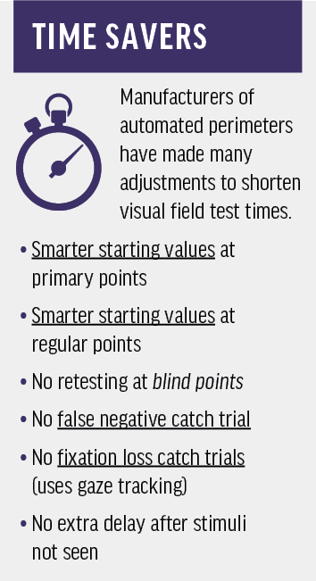
Faster testing options with shorter overall test times have been developed by the major manufacturers of automated perimetry for many years. Initial test times with the original thresholding algorithm averaged around 12 minutes per eye! Over the years, various enhancements have been introduced to shorten the test time (see Time Savers).5
The most recent update, to the best of my knowledge, has been to the Humphrey Field Analyzer 3 (HFA3; Carl Zeiss Meditec), with its SITA Faster program. SITA Faster is another in the SITA line of tests, which include SITA Standard and SITA Fast. It is available to all owners of HFA3 perimeters. Currently, it is capable only of running the 24-2 testing algorithm.
SITA Faster reduces testing time by about half, compared with SITA Standard, and by about one-third compared with SITA Fast. Many patients are able to complete SITA Faster 24-2 testing in about 2 minutes. Clinical testing has shown that SITA Faster produces results that are clinically equivalent to those of SITA Fast with no loss of repeatability.6
An important consideration for those adopting this new program is how to incorporate it for patients who already have a series of older perimetry tests in their history. To facilitate this, the HFA3’s Guided Progression Analysis software has been improved to allow mixing of SITA test strategies. SITA Standard, SITA Fast, and SITA Faster can be freely intermixed in the HFA3’s Guided Progression Analysis. Thus, clinicians can introduce SITA Faster testing without having to establish a new baseline for existing patients.
Overall, SITA Faster is similar to the SITA Fast strategy, but with a significant saving in time. Users of the Octopus perimeter (Haag-Streit) will find the tendency-oriented perimetry to be another quick testing option for patients who are unable to maintain concentration for long periods. However, that strategy may be less sensitive to small localized defects. This should be considered when making clinical decisions.
Published glaucoma guidelines have suggested that some glaucoma patients, with disease that is progressing rapidly despite the patient’s therapeutic regimen, could benefit from more frequent visual field testing to facilitate early detection of changes.7 More frequent testing can also help determine a rate of progression sooner in order to tailor therapy to individual patient needs. A significantly faster testing strategy such as this one may facilitate more frequent visual field testing, thus bringing clinical practice more in line with current recommendations.
STAY UPDATED
Visual field testing is here to stay in the management of patients with glaucoma. It continues to provide vital functional information for the majority of patients. It’s important for practitioners to evaluate their perimeter and its software to determine whether updates or improved statistical analyses are available. If upgrades are available, it is recommended that they be incorporated to ensure the best diagnostic experience for both patient and provider.
- Optometric clinical practice guideline: care of the patient with open angle glaucoma. American Optometric Association. www.aoa.org/documents/optometrists/CPG-9.pdf. Accessed May 16, 2019.
- ICD-10 glaucoma reference guide. American Academy of Ophthalmology and American Glaucoma Society. www.aao.org/Assets/5adb14a6-7e5d-42ea-af51-3db772c4b0c2/636713219263270000/bc-2568-update-icd-10-quick-reference-guides-glaucoma-final-v2-color-pdf?inline=1. Accessed May 29, 2019.
- Kass MA, Heuer DK, Higginbotham EJ, et al. The Ocular Hypertension Treatment Study: a randomized trial determines that topical ocular hypotensive medication delays or prevents the onset of primary open-angle glaucoma. Arch Ophthalmol. 2002;120(6):701-713.
- Kansal V, Armstrong JJ, Pintwala R, Hutnik C. Optical coherence tomography for glaucoma diagnosis: an evidence-based meta-analysis. PLos One. 2018;13(1):e0190621.
- Heijl A, Patella VM, Bengtsson B. The field analyzer primer: effective perimetry. 4th ed. Carl Zeiss Meditec; 2012.
- Heijl A, Patella VM, Chong LX, et al. A new SITA perimetric threshold testing algorithm: construction and a multicenter clinical study. Am J Ophthalmol. 2019;198:154-165.
- Terminology and guidelines for glaucoma. European Glaucoma Society. June 2014. https://bjo.bmj.com/content/bjophthalmol/101/4/1.full.pdf. Accessed May 17, 2019.


