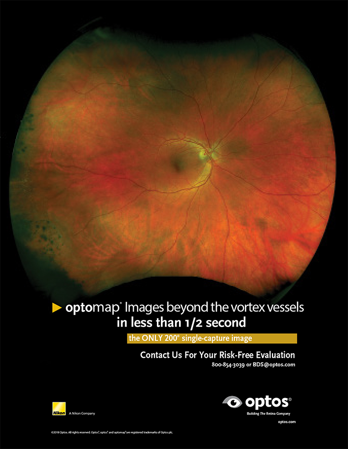

Imaging wide areas of the retina has become substantially easier with the advent of ultrawide-field (UWF) imaging (Figure 1). Images of the periphery can frequently be captured via a single image without pupillary dilation. UWF allows imaging of more than 80% of the retina; older imaging modalities captured only as little as 15% of the retina at a time.
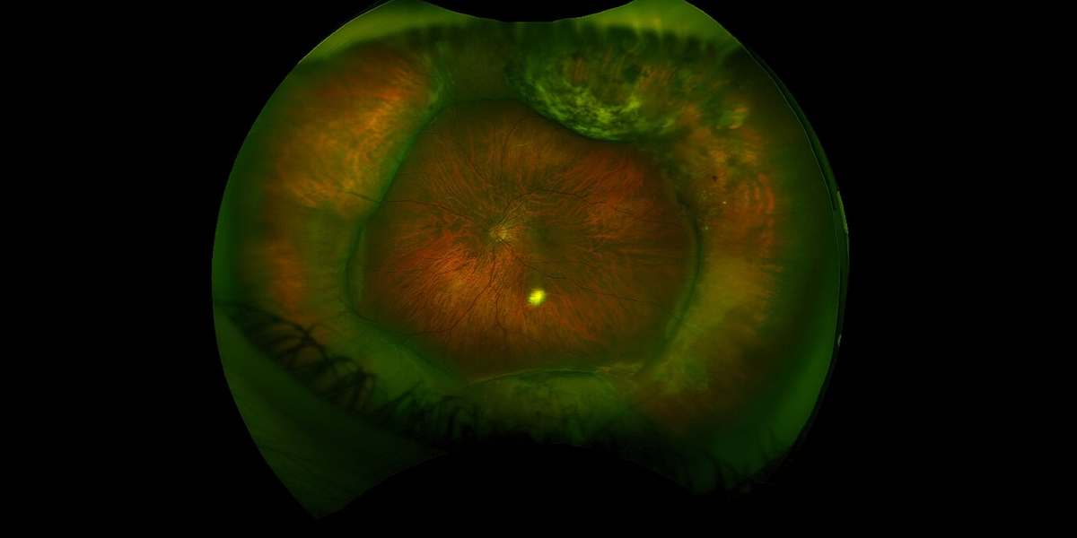
Figure 1. Image of a scleral buckle taken with the Daytona Plus (Optos). Photo credit: Jeffry Gerson, OD, FAAO.
UWF imaging has clinical applications in patients with diabetic retinopathy (DR), age-related macular degeneration, retinal vein occlusion (RVO), and hypertensive retinopathy (Figure 2). The ability to perform UWF fluorescein angiography (FA) in the periphery and evaluate the peripheral fundus for abnormalities such as neovascularization and ischemia may lead to the development of new treatments and treatment algorithms for diseases such as DR.1
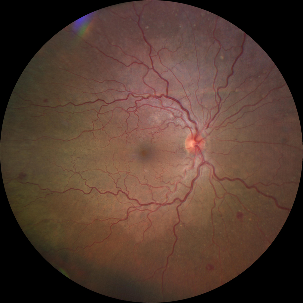
Figure 2. Widefield image of a patient with mild hypertensive retinopathy. Image obtained with the Zeiss Clarus 500 (Carl Zeiss Meditec). Photo credit: Jay Haynie, OD, FAAO.
THE LITERATURE
The literature exploring what UWF imaging can offer to clinicians is robust.
Diabetic Retinopathy
A study comparing UWF imaging with traditional seven-field 45° imaging in diabetic patients found that UWF imaging is easier and more efficient than montaging multiple images for the purpose of capturing a wider image (Figure 3).2 The study found that the two imaging methods had a high degree of agreement. Silva et al showed that that a third of hemorrhage microaneurysms, intraretinal microvascular abnormalities, and new vessels were located outside of the area captured by standard seven-field imaging, resulting in a higher DR grade in 10% of cases on UWF imaging than would have been suggested by seven-field montage.3
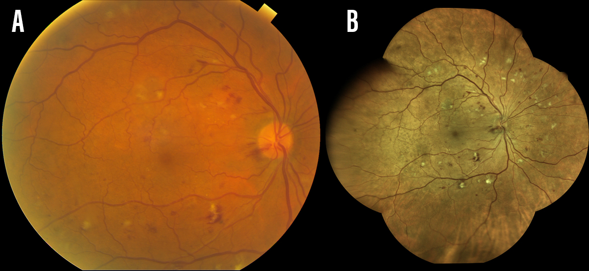
Figure 3. Image of a patient with severe nonproliferative diabetic retinopathy. Image taken with a standard 50° field (A) and in the widefield (B) with the Eidon True Color Confocal Scanner (CenterVue).
Similarly, Wessel et al reported that 10% more retinal neovascularization and retinal nonperfusion was detected with UWF FA than with an overlay of conventional seven standard field imaging.4
Age-Related Macular Degeneration
It has been theorized that peripheral retinal ischemia plays a role in choroidal neovascular membrane formation and may be a risk factor for membrane development. Madhusudhan and Beare found peripheral leakage seen on UWF FA to be associated with the presence of active neovascular age-related macular degeneration.5
Retinal Vein Occlusion
RVO can be imaged and documented with UWF technology (Figure 4). Studies have demonstrated that UWF FA provides visualization of peripheral nonperfusion in patients with branch RVO and hemicentral RVO. This can help clinicians to target areas for panretinal photocoagulation, allowing more efficient treatment of the ischemic retina and potentially less damage to healthy adjacent tissue.6
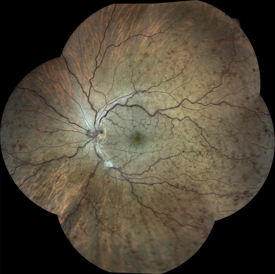
Figure 4. Widefield image of a nonischemic central retinal vein occlusion obtained with the Eidon True Color Confocal Scanner (CenterVue).
WHAT CAN WE SEE?
UWF imaging and UWF FA are valuable tools for understanding the characteristics of retinal pathology. Future technology will undoubtedly improve our ability to visualize the peripheral retina, thereby enhancing our knowledge of various etiologies and aiding in future treatments.

- Tan CS, Sadda SR, Hariprasad SM. Ultra-widefield retinal imaging in the management of diabetic eye diseases. Ophthalmic Surg Lasers Imaging Retina. 2014;45:363-366.
- Rasmussen ML, Broe R, Frydkjaer-Olsen U, et al. Comparison between Early Treatment Diabetic Retinopathy 7-field retinal photos and non-mydriatic, mydriatic, and mydriatic steered widefield scanning laser ophthalmoscopy for assessment of diabetic retinopathy. J Diabetes Complications. 2015;29:99-104.
- Silva PS, Cavallerano JD, Sun JK, et al. Peripheral lesions identified by mydriatic ultrawide field imaging: distribution and potential impact on diabetic retinopathy severity. Ophthalmology. 2013;120(12):2587-2595.
- Wessel MM, Aaker GD, Parlitsis G, et al. Ultra-wide-field angiography improves the detection and classification of diabetic retinopathy. Retina. 2012;32(4):785-791.
- Madhusudhan S, Beare N. Wide-field fluorescein angiography in wet age-related macular degeneration. ScientificWorldJournal. 2014;2014:536161.
- Prasad PS, Oliver SC, Coffee RE, et al. Ultra wide-filed angiographic characteristics of branch retinal and hemicentral retinal vein occlusion. Ophthalmology. 2010;117:780-784.