
In the March/April issue of CollaborativeEYE, I reviewed some recent pharmacologic and surgical innovations that have improved care for patients with diabetic eye disease. Here, I delve into the theme of diabetes care further by exploring imaging innovations that have made it easier for clinicians to detect diabetic retinopathy (DR) and diabetic macular edema (DME).
Specifically, enhanced imaging modalities such as spectral-domain OCT (SD-OCT), widefield imaging, and OCT angiography (OCTA) have given eye care providers a better view of the posterior segment than ever before.
SD-OCT
SD-OCT has become the gold standard in monitoring of DR and DME. Every diabetic patient, regardless of whether he or she is receiving treatment for diabetic eye disease, should have an SD-OCT evaluation at each visit (Figure 1).
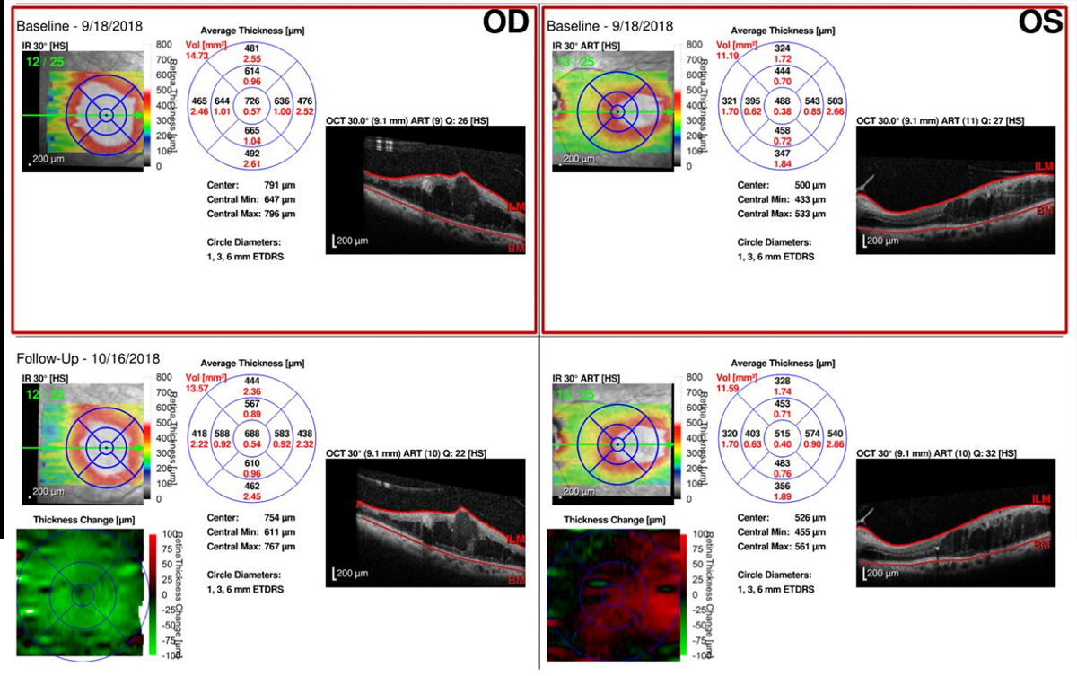
Figure 1. A spectral-domain OCT scan of a patient with diabetic eye disease at baseline (in red boxes) and at 1 month.
SD-OCT allows clinicians to monitor DME and guide treatment decisions, such as whether to adjustment treatment dosage, frequency, or choice of therapeutic agent. Current software provides valuable segmentation algorithms that allow sophisticated progression analysis. One glance shows whether there has been stability, worsening, or improvement in intraretinal and subretinal fluid since the patient’s previous visit, making it an indispensable tool in the clinic.
ULTRAWIDE-FIELD IMAGING
Ultrawide-field imaging continues to provide incredible images of the retina and retinal periphery, even in patients with small pupils. Combining ultrawide-field imaging with fluorescein angiography provides unparalleled evaluation from the macula to the periphery, allowing clinicians to evaluate neovascularization or peripheral capillary nonperfusion (Figure 2).
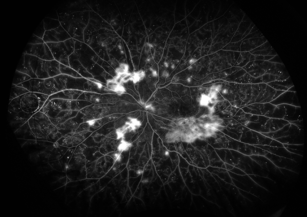
Figure 2. Proliferative diabetic retinopathy as seen on ultawide-field fluorescein angiography.
Ultrawide-field imaging will play an important role in the future of telemedicine screening of diabetic eye disease in at-risk patients. Nikon (with its subsidiary Optos) has already announced a partnership with Alphabet/Verily to develop artificial intelligence technologies to detect retinal diseases based on ultrawide-field retinal images. In April, the FDA approved for marketing the first artificial intelligence device for DR detection, IDx-DR (IDx), a software system that analyzes clinician-uploaded images taken with the NW400 retinal camera (Topcon).
OCTA
OCTA is the most recent innovation in this list. It allows imaging of the retinal vasculature, including the middle and deep vascular plexus and the choriocapillaris. Unlike fluorescein angiography, which requires injection of fluorescein dye that may result in an allergic reaction, OCTA is dyeless. OCTA is currently in investigational stages, but it has already shown significant promise for use in most retinal vascular diseases, including DR. We await further development of the segmentation algorithms, progression analysis, and indications for use.
SEEING IS EVERYTHING
Clinicians who can see a disease at work can better tailor therapy to an individual patient’s needs. Given the variety of imaging platforms eye care providers can now use to detect DR or DME, there is increasing likelihood that patients with early disease can be identified and treated before clinically significant vision is lost.